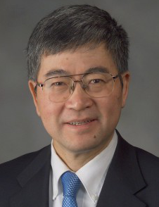
 Biograph: Ge Wang is Clark & Crossan Endowed Chair Professor and Director of Biomedical Imaging Center, Rensselaer Polytechnic Institute, Troy, NY, USA. He published the first spiral/helical cone-beam/multi-slice CT algorithm in 1991 and since then 100+ papers systematically contributed to theory, algorithms, artifact reduction and biomedical applications in this area. Currently, there are 100+ million medical CT scans yearly with a majority in the spiral/helical cone-beam/multi-slice mode. His group developed interior tomography theory and algorithms to solve the long-standing “interior problem” for high-fidelity local reconstruction, and enable omni-tomography (“all-in-one”) with CT-MRI as an example. He initiated the area of bioluminescence tomography. He wrote 450+ journal publications, receiving a high number of citations and academic awards. His results were featured in Nature, Science, PNAS, and various news media. In 2016, he wrote the first perspective on neural-network-based tomographic imaging as the new frontier of machine learning. His team has been in collaboration with world-class groups and continuously well-funded by federal agencies and major imaging companies, actively translating machine learning techniques into imaging products. His interest includes x-ray CT, MRI, optical tomography, multimodality fusion, and machine learning. He is Lead Guest Editor of five IEEE Transactions on Medical Imaging Special Issues, Founding Editor-in-Chief of International Journal of Biomedical Imaging, Outstanding Associate Editor of IEEE Trans. Medical Imaging, Board Member of IEEE Access, Associate Editor of IEEE Trans. Radiation and Plasma Medical Sciences, Medical Physics, and Editorial Board Member of Journal of Machine Learning Science and Technology. He is Fellow of IEEE, SPIE, OSA, AIMBE, AAPM, and AAAS.
Title:Bio-Imaging Driven by Big Data and Deep Learning
Abstract: Currently, deep learning is the mainstream of machine learning and a most active area of artificial intelligence. Computer vision and image analysis are great application examples of deep learning. While computer vision and image analysis deal with existing images and produce related features (registration, segmentation, classification, etc.), tomography produces images of internal structures from externally measured features (line integrals, k-space samples, etc.) of underlying images. Recently, deep learning techniques are being actively developed worldwide for tomographic image reconstruction. We believe that “image reconstruction is a new frontier of machine learning” (IEEE Transactions on Medical Imaging 37 (2018) 1289), and promises major impacts on the development of solutions to many inverse problems. Over the past years, we have been working on data-driven bio-imaging, especially CT, MRI, and optical image reconstruction algorithms for superior imaging performance. In this presentation, we report our representative results, involving important applications and methodological innovations. We welcome collaborative opportunities.